Abdomen with bilateral Hernias
Safety & Quality
Safety & Quality
👉 This product is for training purposes only.
✅ This product is Latex-free.
✅ Sim & Skills is ISO 9001 certified.
✅ Available via NHS Supply Chain.
Delivery
Delivery
When will my order arrive?
Items listed as In Stock are usually delivered in 1-3 working days.
Due to global supply chain disruption, some products are taking longer to deliver. If your order is affected, we will inform you as soon as possible with an estimated lead time or an alternative product recommendation.
For urgent or high volume orders, please contact us before placing your order to check stock availability.
How much does delivery cost?
Tracked Delivery on website orders to a single Mainland Great Britain address is charged at £7.50 + VAT.
Tracked Delivery on purchase orders to a single Mainland Great Britain address is charged at £15.00 + VAT.
Shipping to Northern Ireland and British Forces is charged at £25.00 + VAT.
Palletised deliveries are charged at £85.00 + VAT per pallet.
Deliveries will be made to a Goods In area not a specific room/department.
International shipping is provided on a case-by-case basis, please contact us for a quote.
Returns
Returns
What is the returns policy?
Returns are acceptable within 28 days of delivery (by law you have 14 days to notify us and a further 14 days to complete the return), providing the item is in a resalable condition and in its undamaged original packaging.
Cancellations and Returns are free of charge for items up to 20 kg. Collection for items over 20 kg will be quoted on a case-by-case basis.
All returns must be pre-authorised prior to shipping. Unauthorised returns may be refused and additional carriage costs may be incurred.
Failure to return goods in a resalable condition may result in a restocking fee of £25.00 + VAT or 20% of the invoice value, whichever is greater.
Any goods returned for product defect or warranty issues will not incur a restocking fee, but must still have appropriate authorisation.
To start a return, email orders@simandskills.com.
If your return is accepted, we’ll send you a return shipping label and instructions on how and where to send your package. Items sent back to us without first requesting a return will not be accepted.

Abdomen with bilateral Hernias
Description
Description
Abdomen with bilateral Hernias
This 3D model represents one of the largest and most complex in the series, consisting of a partial torso from the diaphragm to the proximal thigh with a complete abdominal cavity preserving varying levels of dissection. This 3D model also records the rare, simultaneous occurrence of indirect and direct inguinal hernias allowing for a consideration of the anatomical underpinnings for both conditions. Given the scale of the dissection this 3D model description is divided into discrete parts based on views and regions.
The diaphragm
The diaphragm is preserved on the model’s superior aspect, with both domes and costodiaphragmatic recesses visible despite some distortion from rib removal. The fibrous pericardium rests on the central tendon, with the terminal inferior vena cava seen in the caval foramen. Lateral to this lies the oesophagus in the oesophageal hiatus, and the descending thoracic aorta approaching the aortic hiatus near the vertebrae.
The epigastric and hypochondriac regions
In the abdomen, removal of the anterior wall, greater omentum, and much of the GI tract reveals retroperitoneal structures. The terminal oesophagus enters just left of the liver. With the stomach removed, the pancreas is fully exposed from head to tail, reaching the spleen in the left hypochondrium. Above it, the splenic and common hepatic arteries span the narrow space between pancreas, diaphragm, and liver. The tortuous splenic artery divides near the splenic vein; the common hepatic gives rise to the gastroduodenal and right gastric arteries, superficial to the portal vein. The superior mesenteric vessels pass near the pancreatic head, and the ileocolic artery leads to the caecum. The inferior mesenteric vein arises from the superior rectal vein and crosses the descending aorta.
Below the liver, the gallbladder lies between the lobes. On the left, renal vessels pass deep to the pancreas, with ureters descending across the psoas muscles.
The umbilical and lumbar regions
Most abdominal organs in the umbilical and lumbar regions have been removed to reveal the posterior abdominal wall. Centrally, the descending aorta and inferior vena cava are prominent, with testicular vessels traceable toward the inguinal region. Two right lumbar arteries branch from the aorta, and the inferior mesenteric artery gives rise to the left colic, sigmoid, and superior rectal arteries. On the right, subcostal, iliohypogastric, and ilioinguinal nerves are visible, along with the circumflex iliac artery.
The hypogastrium and iliac regions
The abdominal aorta bifurcates into the common, internal, and external iliac arteries, with matching iliac veins merging into the inferior vena cava. The obturator artery, ureters, and testicular vessels are visible. In the true pelvis, the peritoneum covers the bladder, while the rectum remains obscured. The right iliac fossa contains the terminal ileum, caecum, and appendix, with nearby vessels and nerves. On the left, the sigmoid colon crosses the iliac fossa, where an epiploic appendage extends into an indirect hernia near the inferior epigastric artery.
The inguinal region and perineum
This model uniquely preserves both direct (right) and indirect (left) inguinal hernias, with the inferior epigastric vessels retained for anatomical orientation. The right hernia lies medial to these vessels; the left hernia sac extends laterally into the spermatic cord, containing an epiploic appendage. The perineum reveals the penis, testes, and spermatic cords. On the right, the cord remains intact; on the left, it’s opened, showing a varicose testicular vein linked to the indirect hernia.
The thigh
The femoral triangle has been dissected on both thighs. On the right, the femoral sheath was removed to reveal the femoral artery, vein, deep inguinal lymph nodes, and femoral nerve. On the left, a broader view exposes anterior and medial thigh muscles, with the femoral artery, profunda femoris, and circumflex iliac artery visible. The model ends mid-thigh, showing cross-sectional anatomy including the femoral shaft, vessels, and muscles in the subsartorial canal.
Specifications
Specifications
-
Weight
-
Manufacturer's Warranty1 Year
-
Downloads
Reviews (0)
Reviews (0)
Payment & Security
Secure payment methods
Your payment information is processed securely. We do not store or have access to your credit card details.
Purchase Orders
We accept purchase orders from public and government authorities including the NHS and Universities subject to our terms of business.
Click Add to Quote to easily build your quote ➡️
Let customers speak for us

Very life like ,easy to use

The quality is amazing 🥰
Amazing
But recommend the sutures to be 2/0.
4/0 is very small
I'm a medical student and was really pleased with the quality of this kit, all the tools are well made and the case is near and sturdy

Works well
If i could give it 4.5 I would but I'm much more inclined to say I'm happy with this product. Really good quality, excellent resource for my vet suturing practice. I have a mild complaint about the silk sutures, they rip/ fray more easily than I hoped, but easily rectified by buying new ones (which are cheap) and also encourages me to be more gentle with my knots. I'm so happy with it and it's a perfect still revising break from studying. Thank you Sim and Skills!!!

Student Feedback: Feedback was gathered from 13 students. Overall, they liked the trainer and would recommend it to their peers for learning blood glucose testing. They found the trainer easy to use and helpful in demonstrating the procedure. The students felt that the trainer added significant value to their training, with the main benefit being an increased competency in blood glucose testing.
Lecturer Feedback: Feedback was collected from 7 lecturers. Most of them found the trainer to be authentic and effective in simulating the challenges of blood sugar testing. They appreciated that it encouraged patient communication, which is a great plus. The product was of good quality and reliable for teaching purposes. However, there was a minor issue with the finger becoming leaky after multiple uses. Despite this, the lecturers agreed that the trainer prepares students well for real-world blood glucose testing.

Excellent service and speedy delivery. Highly recommended
Brilliant teaching aid. Lots of useful equipment within set. Excellent for practice.
Amazing product

As a proud owner of a Sim & Skills Foot & Ankle model, I thought it was time I invested in a Hand & Wrist.
My clients find it really helpful when we are discussing anatomy and it really aids their recovery.
I believe client education and knowing that your body is something to be enjoyed, not feared, is such an important platform for recovery from chronic pain.

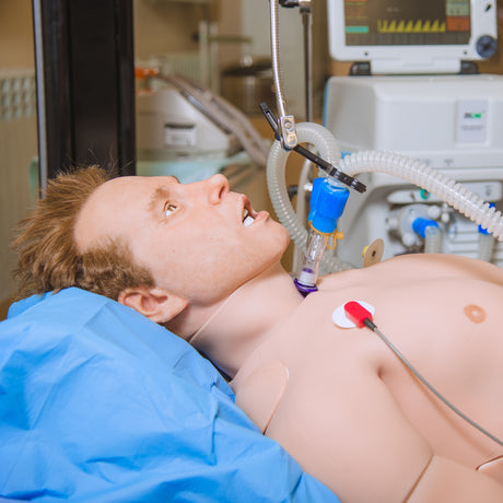
This item is very useful for NG insertion training, it can be used in a horizontal position and the fluid bag does not leak out. The bag and tubing can all be stored within the head making storage of the item easy. It is very simple to use and the head is strong and durable
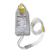
Purchased these bags for training with venepuncture and so far very pleased with quality and cost

Fast delivery, excellent communication and a great price

Excellent fast service. Thank you.