The use of the central venous catheter (CVC) or Central Line insertion, has become an invaluable resuscitation tool in intensive care units and A&E departments.
What are the risks of a Central Line?However, Central line placement has risks, including the potential for infection in patients if inserted incorrectly.
Experience with Central Line insertion has often come solely from patient encounters, which can lead to potential risks for the patient. Previous studies have shown that up to 15% of patients have experienced CVC insertion complications
How can Central Line complications be prevented?Experiential learning using Central Line placement training models, can help to limit these risks and improve patient safety.
A study published in the Journal of the Society for Simulation in Healthcare found that ultrasound guided training led to an increase in a clinician’s proficiency, with improved Central Line placement.
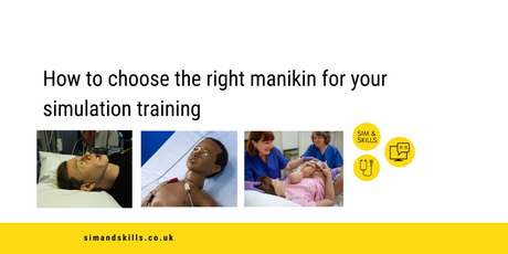
Here are the Top 5 reasons why you should be using Blue Phantom’s Gen II Central Line Ultrasound training model
-
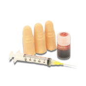
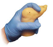
It mimics soft human tissue and vascular structures, allowing ultrasound learners to train in needle-guided procedures without exposing actual patients to the pain and discomfort that is often associated with repeated attempts at proper needle insertion.

-
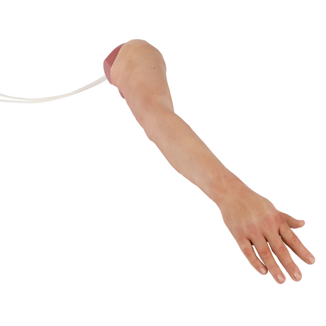
The upper torso models are ideal for practising both ultrasound-guided and blind central line placement. This model is excellent for using traditional external landmarks for blind insertion central line placement, or ultrasound to guide the central venous access procedure.

-
The simulated skin tissue will "self-heal." It will automatically seal itself around the puncture area, eliminating the risk of leakage and reducing the appearance of damage.

-
There are 4 options to choose from: Opaque Insert with Auto Pump, Opaque Insert with Hand Pump, Transparent Insert with Auto Pump, or Transparent Insert with Hand Pump.

- The Blue Phantom task trainer model can endure 1,000 needle attempts or more, without significant degradation in the integrity of the simulated training model, the simulated vessel, or the ultrasound image produced.