Abscess Drainage Ultrasound Training Model
£1,75200Unit price /Unavailable

Amniocentesis Ultrasound Training Model
£11,98800Unit price /Unavailable

Bone Fracture Ultrasound Training Block Model
£1,75200Unit price /Unavailable-

Branched 2 Vessel Ultrasound Training Block Model
£87600Unit price /Unavailable 
 BEST SELLER
Branched 4 Vessel Ultrasound Training Block Model
£1,02000Unit price /Unavailable

Breast Biopsy Ultrasound Training Model
£87600Unit price /Unavailable

Combination IUP Ectopic Pregnancy Transvaginal Ultrasound Training Model
£14,77200Unit price /Unavailable

Elastography Ultrasound Breast Phantom
£87600Unit price /Unavailable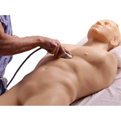
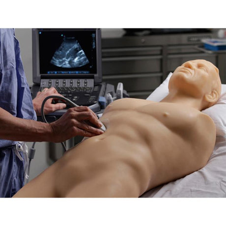
FAST Exam Ultrasound Training Model
From £47,22000Unit price /Unavailable-

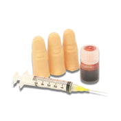
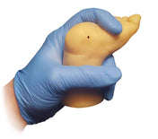
Foreign Body Identification Ultrasound Training Model
£1,02000Unit price /Unavailable 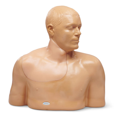
 BEST SELLER
Gen II Central Line Ultrasound Training Model
From £4,68000Unit price /Unavailable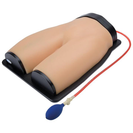
 BEST SELLER
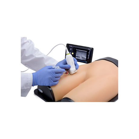
Gen II Femoral Vascular Access and Regional Anaesthesia Ultrasound Training Model
£7,30800Unit price /Unavailable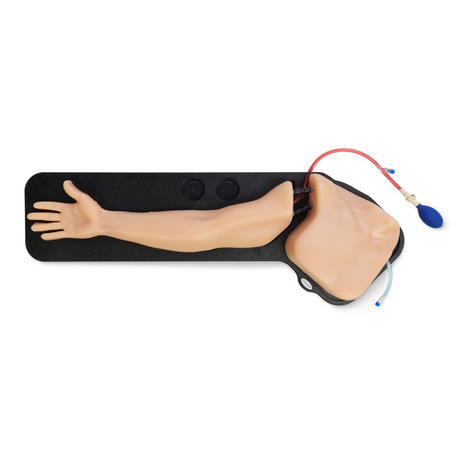
 BEST SELLER
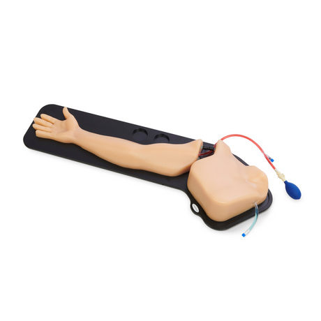
Gen II PICC with IV & Arterial Line Vascular Access Ultrasound Trainer
£4,68000Unit price /Unavailable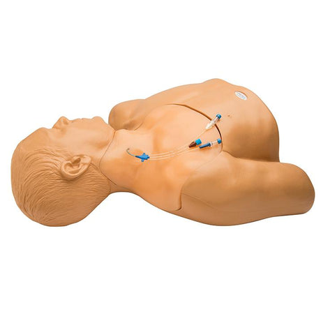
 BEST SELLER
Gen II Regional Anaesthesia and Central Line Ultrasound Training Model
£10,96800Unit price /Unavailable

General Pathology Transvaginal Ultrasound Training Model
£12,57600Unit price /Unavailable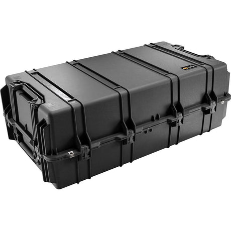
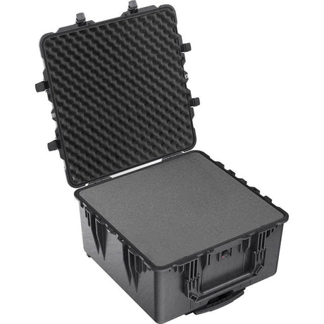
Hard Case for FAST and Echocardiography Full Torsos
£2,34000Unit price /Unavailable

Hard Case for Gen II Central Line Ultrasound Training Model
£72000Unit price /Unavailable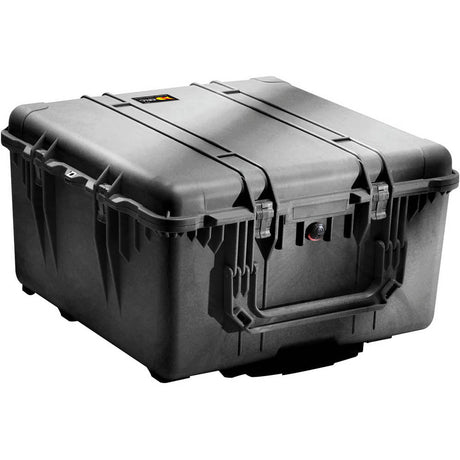
Hard Case for Gen II Femoral Vascular Access and Regional Anaesthesia Ultrasound Training Model
£72000Unit price /Unavailable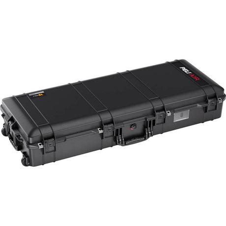
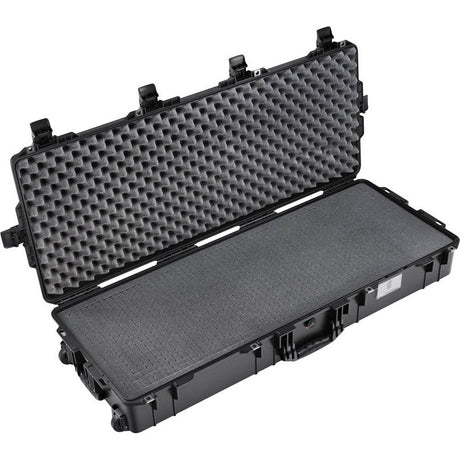
Hard Case for Gen II PICC with IV & Arterial Line Vascular Access Ultrasound Trainer
£72000Unit price /Unavailable


Internal Jugular Central Line Ultrasound Manikin - Transparent/Flesh
From £3,14400Unit price /Unavailable